Imaging Analysis & Live Cell Imaging

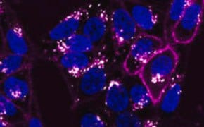
Live-cell imaging and analysis allows scientists to study dynamic cellular events in real time to gain unique insights into biology. These techniques are in contrast to PCR, flow cytometry, and immunocytochemistry and tissue staining with antibodies, which analyze cellular events as a snapshot. Today, improvements in cameras, live-cell fluorescent dyes, fluorescent proteins, video compression, and time-lapse microscopy have advanced our ability to visualize and analyze living cells, their intercellular interactions, and subcellular processes with remarkable detail and fidelity.
Featured Categories
Duolink® Proximity Ligation Assay (PLA) technology for detecting and quantifying...
Explore our assays for mimicking cell behaviors in vivo. Migration, invasion, Boyden chamber, and...
The CellASIC® ONIX2 Microfluidic System is a powerful, automated platform that allows precise manipulation of the cell culture environment for live-cell imaging and analysis using mammalian, yeast, and bacterial cells.
Standard, complex, and specialty cell culture media for a variety of cell lines and applications. We also offer starting buffers and supplements used in custom media development.
Live-cell Imaging Systems
A variety of techniques are used to visualize the complex structures and behavior of cells. In static analyses, cells are fixed and stained, which requires killing the cells and therefore capturing a single instant in cellular time. While static techniques are suitable for some applications, they do not provide insight into cellular activities. Dynamic, live-cell approaches facilitate the visualization of structure, behavior or composition in live cells, without traditional staining. These techniques are ideal for applications that must mimic in vivo conditions, as well as for developing predictive assays for drug screening.
Live-cell imaging systems use advanced optics, environmental control, and reagents that permit long-term studies of mammalian, bacterial, and yeast cells. Culture and imaging systems that also employ microfluidics allow precise control of the culture microenvironment, like flow, gas mixture, and temperature, while limiting physical stress to cells. Live-cell imaging and analysis is suited to a range of applications, including hypoxia, cell migration, 3D cell culture, cytoskeletal dynamics and protein trafficking. Advanced systems automate culture conditions, administration and withdrawal of treatments, and imaging intervals to create images that may be presented as still image or video data.
Live-cell Imaging Reagents
Live-cell imaging media and supplements are designed to protect cells from light-induced cellular damage during time-lapse experiments. These reagents are also formulated to have low autofluorescence and photobleaching, which dramatically enhances the quality of fluorescent live-cell images.
A variety of live-cell staining dyes are available to study the dynamic processes of live cells. Live-cell fluorescent organelle dyes permit the selective staining of specific organelles, such as the cell membrane, nucleus, cytoplasm, mitochondria, lysosomes, endoplasmic reticulum (ER), Golgi apparatus, and cytoskeleton proteins, all without increased cytotoxicity. Using organelle dyes as counterstains in live-cell imaging is also useful in functional studies.
Biosensors with GFP and RFP can be used to detect a specific protein as well as the subcellular location of that protein within live cells by either fluorescent microscopy or time-lapse video capture. Lentiviral vector systems are a popular tool for introducing genes and gene products into cells, with many advantages over chemical-based transfection. Beyond organelle detection, cell-permeable live-cell dyes are used for applications such as apoptosis detection, cell viability, cell health, hypoxia, reactive oxygen species studies, calcium indicator imaging, and neural or stem cell experiments.
Looking for More Specific Information?
Visit our document library for data sheets, certificates and technical documentation.
Related Articles
- Cell based assays for cell proliferation (BrdU, MTT, WST1), cell viability and cytotoxicity experiments for applications in cancer, neuroscience and stem cell research.
- Firefly luciferase is a sensitive reporter for gene studies due to its absence in mammalian cells or tissues.
- PKH and CellVue® Fluorescent Cell Linker Kits provide fluorescent labeling of live cells over an extended period of time, with no apparent toxic effects.
- Fluorescent calcium indicators and dyes to measure Ca+ flux used in calcium imaging experiments. Calcium ions (Ca2+) play vital cellular physiology roles in signal transduction pathways, in neurotransmitter release, in contraction of all muscle cell types, as enzyme cofactors, and in fertilization.
- Lipophilic cell tracking dyes enable cancer biologists to track tumor and immune cell functions both in vitro and in vivo. Read the article to choose a right membrane dye kit for cell tracking and proliferation monitoring.
- See All (29)
Related Protocols
- PKH dyes label exosomes for tracking experiments in vitro and in vivo, detailed protocol provided.
- Cell staining can be divided into four steps: cell preparation, fixation, application of antibody, and evaluation.
- ICC Cell Capture Imaging Reagent simplifies imaging, ideal for rare cell and low cell count samples.
- Toluidine blue selectively stains nuclear material and acidic tissue components, aiding in histological staining for tissues rich in DNA/RNA.
- This protocol covers 3 modes for the microscopic examination of cell samples.
- See All (6)
Find More Articles and Protocols
How Can We Help
In case of any questions, please submit a customer support request
or talk to our customer service team:
Email [email protected]
or call +1 (800) 244-1173
Additional Support
- Chromatogram Search
Use the Chromatogram Search to identify unknown compounds in your sample.
- Calculators & Apps
Web Toolbox - science research tools and resources for analytical chemistry, life science, chemical synthesis and materials science.
- Customer Support Request
Customer support including help with orders, products, accounts, and website technical issues.
- FAQ
Explore our Frequently Asked Questions for answers to commonly asked questions about our products and services.
To continue reading please sign in or create an account.
Don't Have An Account?


