
Z-2183

ZytoLight® SPEC EWSR1/FLI1 TriCheck Probe
Green/Orange/Blue, pkg of 0.05 mL
Synonym(s):
Dual color probe, fluorochrome, probe
Sign In to View Organizational & Contract Pricing.
Select a Size
Change View
| Size/SKU | Availability | Price |
|---|
About This Item
eCl@ss:
32150313
Product Z-2183 is not currently sold in your country. Contact Technical Service
product line
ZytoVision
packaging
pkg of 0.05 mL, pkg of 5 tests
manufacturer/tradename
ZytoVision
storage condition
protect from light
color
, Green/Orange/Blue
application(s)
research use
storage temp.
2-8°C
General description
The ZytoLight®SPEC EWSR1/FLI1 TriCheck Probe is composed of:
- ZyGreen (excitation 503 nm/emission528 nm) labeled polynucleotides (~10ng/µl), which target sequences mappingin 22q12.2** (chr22:29,779,841-30,179,900) distal to the EWSR1 breakpoint region
- ZyOrange (excitation 547 nm/emission 572 nm) labeled polynucleotides(~4.5 ng/µl), which target sequences mapping in 22q12.1-q12.2**(chr22:29,191,431-29,673,440) proximal to the EWSR1 breakpoint region
- ZyBlue (excitation at 418 nm andemission 467 nm) labeled polynucleotides (~37 ng/µl), which target sequences mapping in 11q24.3**(chr11:128,707,454-129,346,602) distal to the FLI1 breakpoint region
- Formamide based hybridization buffer
The fluorescence in situ hybridization (FISH) technique allows for the detection and visualization of specific nucleic acid sequences in cell preparations. Following DAPI counterstaining of the DNA, hybridized probe fragments are observed using a fluorescence microscope with filters tailored for the fluorochromes is used to label the FISH probe fragments.
The ZytoLight® SPEC EWSR1/FLI1 TriCheck Probe (PL141) is a probe for for the qualitative detection of rearrangements involving the humanEWSR1 gene and the human FLI1 gene
Application
- Differentiating Ewing sarcoma/PNET from classic neuroblastoma, Wilms tumor, and rhabdomyosarcoma is crucial.
- The t(11;22)(q24;q12.2) translocation, which juxtaposes the EWSR1 gene at 22q12.2 next to the FLI1 locus, is the most frequent translocation involving the EWSR1 gene region.
- Translocations in the 22q12.2 region are found in 90-95% of patients with Ewing sarcoma
- Therefore, the ZytoLight® SPEC EWSR1/FLI1 TriCheck Probe is used to detect EWSR1 rearrangements by fluorescence in situ hybridization (FISH) to confirm the presence of Ewing sarcoma/PNET.
Analysis Note
- In an interphase nucleus without a FLI1-EWSR1 rearrangement, two green/orange fusion signals and two blue signals are expected.
- A FLI1-EWSR1 fusion is indicated by:
- One separate orange signal co-localizing with one blue signal.
- One separate green signal.
- An EWSR1 translocation not involving FLI1 is indicated by:
- The separation of one green/orange fusion signal.
- No co-localization of the separated orange signal with one blue signal.
Legal Information
ZytoLight is a registered trademark of ZytoVision GmbH in Germany
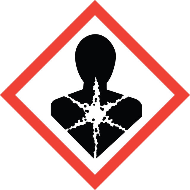
Signal Word
Danger
Hazard Codes
Precautionary Statements
Hazard Classifications
Carc. 2 - Repr. 1B - STOT RE 2 Oral
Target Organs
Blood
Storage Class
6.1C - Combustible acute toxic Cat.3 / toxic compounds or compounds which causing chronic effects
Regulatory Listings
Regulatory Listings are mainly provided for chemical products. Only limited information can be provided here for non-chemical products. No entry means none of the components are listed. It is the user’s obligation to ensure the safe and legal use of the product.
75-12-7
CAS No.
75-12-7
CAS No.
Choose from one of the most recent versions:
Already Own This Product?
Find documentation for the products that you have recently purchased in the Document Library.