


The protocol is published in the product datasheet, available in the 'DOCUMENTATION' section of this page, under the 'Mor Documents' tab. Please see the link below to review:
https://www.sigmaaldrich.com/deepweb/assets/sigmaaldrich/product/documents/271/475/mini26bul.pdf
MINI26
PKH26 Red Fluorescent Cell Linker Mini Kit for General Cell Membrane Labeling
Distributed for Phanos Technologies
Synonym(s):
Red PKH membrane label kit
Sign In to View Organizational & Contract Pricing
Select a Size
1 KIT
€384.00
€384.00
Please contact Customer Service for Availability
About This Item
UNSPSC Code:
12352207
NACRES:
NA.32
Skip To
packaging
pkg of 1 kit
manufacturer/tradename
Distributed for Phanos Technologies
storage condition
protect from light
technique(s)
flow cytometry: suitable
fluorescence
λex 551 nm; λem 567 nm (PKH26 dye)
application(s)
cell analysis
detection
detection method
fluorometric
shipped in
ambient
storage temp.
room temp
1 of 4
This Item | PKH26GL | MINI67 | PKH26PCL |
|---|---|---|---|
| technique(s) flow cytometry: suitable | technique(s) - | technique(s) flow cytometry: suitable | technique(s) - |
| packaging pkg of 1 kit | packaging pkg of 1 kit | packaging pkg of 1 kit | packaging - |
| manufacturer/tradename Distributed for Phanos Technologies | manufacturer/tradename Distributed for Phanos Technologies | manufacturer/tradename Distributed for Phanos Technologies | manufacturer/tradename - |
| storage condition protect from light | storage condition protect from light | storage condition protect from light | storage condition - |
| fluorescence λex 551 nm; λem 567 nm (PKH26 dye) | fluorescence λex 551 nm; λem 567 nm (PKH26 dye) | fluorescence λex 490 nm; λem 502 nm (PKH67 dye) | fluorescence - |
| application(s) cell analysis | application(s) - | application(s) cell analysis | application(s) - |
General description
This kit provides enough dye for up to 25 samples of 20 million cells each and enough diluent for up to 5 samples of 20 million cells each, when using a 2 mL final staining volume containing a final concentration of 2 x 10⁻⁶ M of PKH26 dye. Users must determine the optimal dye and cell concentrations from their cell type(s) and experimental purposes.
Application
PKH26 red fluorescent cell linker mini kit has been used for:
Other Notes
For additional technical details on PKH and CellVue® Fluorescent Cell Linker Dyes including an extensive bibliography, please visit here.
Legal Information
CellVue is a registered trademark of Phanos Technologies
Kit Components Only
Product No.
Description
- Diluent C 10 mL
- PKH26 Cell Linker in ethanol .1 mL
Signal Word
Danger
Hazard Statements
Precautionary Statements
Hazard Classifications
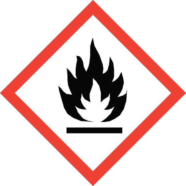
Eye Irrit. 2 - Flam. Liq. 2
Storage Class Code
3 - Flammable liquids
Flash Point(F)
57.2 °F - closed cup
Flash Point(C)
14 °C - closed cup
Choose from one of the most recent versions:
Already Own This Product?
Find documentation for the products that you have recently purchased in the Document Library.
Combined use of decellularized allogeneic artery conduits with autologous transdifferentiated adipose-derived stem cells for facial nerve regeneration in rats
Sun F, et al.
Biomaterials, 32(32), 8118-8128 (2011)
Transplanted bone marrow stem cells relocate to infarct penumbra and co-express endogenous proliferative and immature neuronal markers in a mouse model of ischemic cerebral stroke
Zhang XM, et al.
BMC Neuroscience, 11(1), 138-138 (2010)
Rakesh Arya et al.
Proteomics. Clinical applications, 14(1), e1900062-e1900062 (2019-09-19)
Detailed understanding of host pathogen interaction in tuberculosis is an important avenue for identifying novel therapeutic targets. Small extracellular vesicles (EVs) like exosomes that are rich in proteins, nucleic acids and lipids, act as messengers and may show altered composition
Lars T van der Veken et al.
Journal of immunology (Baltimore, Md. : 1950), 182(1), 92-101 (2008-12-26)
Killer Ig-like receptors (KIR) are expressed by human NK cells and T cells. Although Ag-specific cytolytic activity and cytokine production of KIR(+) T cells can be inhibited by KIR ligation, the effect of KIR on proliferation is unclear. KIR(+) T
Pallavi V Raja Manuri et al.
Human gene therapy, 21(4), 427-437 (2009-11-13)
Nonviral integrating vectors can be used for expression of therapeutic genes. piggyBac (PB), a transposon/transposase system, has been used to efficiently generate induced pluripotent stems cells from somatic cells, without genetic alteration. In this paper, we apply PB transposition to
Articles
Lipophilic cell tracking dyes enable cancer biologists to track tumor and immune cell functions both in vitro and in vivo. Read the article to choose a right membrane dye kit for cell tracking and proliferation monitoring.
Optimal staining is a key component for studying tumorigenesis and progression. Learn useful tips and techniques for dye applications, including examples from recent studies.
PKH and CellVue® Fluorescent Cell Linker Kits provide fluorescent labeling of live cells over an extended period of time, with no apparent toxic effects.
PKH dyes are easy to use and achieve stable, uniform, and reproducible fluorescent labeling of live cells. PKH dyes are non-toxic membrane stains which produce high signal to noise ratio.
-
MINI26-1KTの実験プロトコルを入手したいのですが、どこにありますか?
1 answer-
Helpful?
-
-
Is it possible to lable cells with PKH26 at 4oC?
1 answer-
The membrane dyes will remain stable during the monoclonal staining at 4 deg C; however, capping of the monoclonal antibodies is highly probable if the general cell membrane labeling is carried out at ambient temperature subsequent to antibody labeling. As additional information may be required to address the question completely, please navigate to the link https://www.sigmaaldrich.com/techservice, click on "Product Technical Inquiries" under the Products Section with all the information needed so that a member of the Technical Service team can reach out to assist further.
For more information, please refer to the product data-sheet below: https://www.sigmaaldrich.com/deepweb/assets/sigmaaldrich/product/documents/271/475/mini26bul.pdf.
Helpful?
-
Active Filters
Our team of scientists has experience in all areas of research including Life Science, Material Science, Chemical Synthesis, Chromatography, Analytical and many others.
Contact Technical Service