
Bone tissue is identified by the green-stained collagen-rich bone matrix and osteoid. Nuclei appear dark brown to black; cytoplasm and muscle stain brick red to bright red; erythrocytes appear bright orange. Bone identification relies on green-stained matrix plus bone morphology (trabecular/lamellar structure) and osteocytes in lacunae. Please see the link below to review a publication that may be helpful:
https://pmc.ncbi.nlm.nih.gov/articles/PMC3979890/
1.00485

Masson-Goldner-Färbekit
for the visualization of connective tissue with trichromic staining
Anmelden zur Ansicht der Organisations- und Vertragspreise.
Größe auswählen
Ansicht ändern
| Größe/SKU | Verfügbarkeit | Preis |
|---|---|---|
500 x 4 mL | Warenkorb auf Verfügbarkeit prüfen | € 527,00 |
Über diesen Artikel
NACRES:
NA.41
UNSPSC Code:
41116124
Technischer Dienst
Benötigen Sie Hilfe? Unser Team von erfahrenen Wissenschaftlern ist für Sie da.
Unterstützung erhaltenQuality Segment
IVD
for in vitro diagnostic use
dilution
(for the visualization of connective tissue with trichromic staining)
application(s)
clinical testing
diagnostic assay manufacturing
hematology
histology
storage temp.
15-25°C
General description
Das Masson-Goldner-Färbungskit für die Veranschaulichung von Bindegewebe mit Trichromfärbung wird für die humanmedizinische Zelldiagnostik verwendet und dient dem Zweck der histologischen Untersuchung von Probenmaterial menschlichen Ursprungs. Durch eine Kombination von drei verschiedenen Färbungslösungen können Muskelfasern, Kollagenfasern, Fibrin und Erythrozyten selektiv veranschaulicht werden.
Die ursprünglichen Verfahren wurden primär verwendet, um Kollagen- und Muskelfasern zu differenzieren. Die verwendeten Farbstoffe weisen unterschiedliche Molekülgrößen auf und ermöglichen, dass verschiedene Gewebe unterschiedlich gefärbt werden.
Die Masson-Goldner-Färbetechnik kann mit formalinfixiertem Material durchgeführt werden. Nach dem Färben des Kerns mit Weigerts Eisenhämatoxylin werden Komponenten wie Muskeln, Zytoplasma und Erythrozyten mit Azophloxin und Orange G-Lösung gefärbt. Anschließend wird Bindegewebe mit hellgrüner SF-Lösung gegengefärbt.
Die Packung reicht für 400 bis 500 Anwendungen. Das Produkt ist ein IVD-Produkt mit CE-Kennzeichnung und kann in der Diagnostik und in Laborakkreditierungen verwendet werden. Weitere Details finden Sie in der Gebrauchsanweisung. Diese Gebrauchsanweisung kann auf dieser Website heruntergeladen werden.
Die ursprünglichen Verfahren wurden primär verwendet, um Kollagen- und Muskelfasern zu differenzieren. Die verwendeten Farbstoffe weisen unterschiedliche Molekülgrößen auf und ermöglichen, dass verschiedene Gewebe unterschiedlich gefärbt werden.
Die Masson-Goldner-Färbetechnik kann mit formalinfixiertem Material durchgeführt werden. Nach dem Färben des Kerns mit Weigerts Eisenhämatoxylin werden Komponenten wie Muskeln, Zytoplasma und Erythrozyten mit Azophloxin und Orange G-Lösung gefärbt. Anschließend wird Bindegewebe mit hellgrüner SF-Lösung gegengefärbt.
Die Packung reicht für 400 bis 500 Anwendungen. Das Produkt ist ein IVD-Produkt mit CE-Kennzeichnung und kann in der Diagnostik und in Laborakkreditierungen verwendet werden. Weitere Details finden Sie in der Gebrauchsanweisung. Diese Gebrauchsanweisung kann auf dieser Website heruntergeladen werden.
Analysis Note
Eignung für Mikroskopie (Gewebeschnitt): entspricht
Kerne: dunkelbraun bis schwarz
Zytoplasma: ziegelrot
Muskulatur (Muskeln): hellrot
Bindegewebe: grün
saure Mukosubstanzen: grün
Erythrozyten: hell orange
Kerne: dunkelbraun bis schwarz
Zytoplasma: ziegelrot
Muskulatur (Muskeln): hellrot
Bindegewebe: grün
saure Mukosubstanzen: grün
Erythrozyten: hell orange
Signalwort
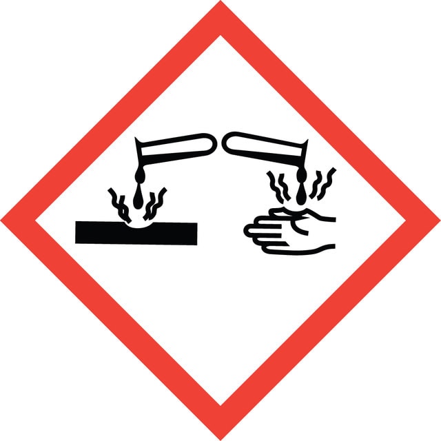
Danger
Was ist los?
WGK 2
Gefahrencodes
Sicherheitshinweise
Hazard Classifications
Aquatic Chronic 3 - Eye Dam. 1 - Skin Irrit. 2
Lagerklasse
8B - Non-combustible corrosive hazardous materials
Hier finden Sie alle aktuellen Versionen:
Besitzen Sie dieses Produkt bereits?
In der Dokumentenbibliothek finden Sie die Dokumentation zu den Produkten, die Sie kürzlich erworben haben.
-
how to identify bone tissue in Masson-golderner stain?
1 answer-
Helpful?
-
-
May I ask if Goldner’s Trichrome and Masson–Goldner are the same staining method? I am planning to use it for undecalcified resin-embedded hard tissue sections to observe newly regenerated bone.
1 answer-
No. The Masson Goldner Trichrome procedure is a modification of the Goldner Trichrome method for mineralized bone. The Masson Goldner Trichrome kit is intended for use with paraffin-embedded tissue to visualize connective tissue. No procedure is provided for staining resin embedded sections of bone. In the Masson Golder staining kit, the deparaffinized tissue sections are stained in Weigert's Iron Hematoxylin for only 5 minutes. In a standard Goldner Trichrome method the sections are stained in Weigert's Iron Hematoxylin solution for 1 hour. Other steps in the procedure also require longer staining times.
Helpful?
-
-
Can I use this kit to visualize fibrosis?
1 answer-
The Goldner Trichrome kit can be one of the tests performed to aid in the diagnosis of various types of fibrosis. Blood testing is sometimes used to rule out other diseases. Imaging tests may be used if pulmonary fibrosis is expected. A hematoxylin and eosin stain may be performed initially.
Fibrosis is a disease where interstitial tissue overgrows, hardens, and scars. This leads to chronic inflammation. The various Trichrome methods (Goldner, Goldner Masson, Gomori and Masson) can be used. The various trichrome methods typically identify muscle and collagen. Interstitial tissue is composed of collagen, elastin, and glycosaminoglycans. The Goldner Masson Trichrome procedure also identifies fibrin and red blood cells.
Helpful?
-
-
Hello, I wonder what is the difference of theTrichrome Stain (Masson) Kit (HT15) and Masson-Goldner staining kit (1.00485.0001)? Thanks
1 answer-
The 1.00485.0001, Masson-Goldner Kit, is primarily used for the visualization of connective tissue with trichromic staining. This kit Includes a phosphotungstate reagent, a light green dye to stain collagen/connective tissues, and Azophloxine(as described by Goldner).
The HT15, Trichrome (Masson) Kit, is intended for use in the study of connective tissue, muscle, and collagen fibers. This kit includes both, a phosphotungstate and a phosphomolybdate component, Aniline blue to specifically stain collagen, and Biebrich Scarlet – Acid Fuchsin.
Both kits require the use of Weigert's Iron Hematoxylin, which is not included.For a more comprehensive understanding of the kits, please see the links below to review the product datasheets:
HT15 Trichrome (Masson) kit
https://www.sigmaaldrich.com/deepweb/assets/sigmaaldrich/product/documents/274/001/procedure-ht15ug-ms.pdf1.00485.0001 Masson-Goldner kit
https://www.sigmaaldrich.com/deepweb/assets/sigmaaldrich/product/documents/124/226/100485-en.pdfHelpful?
-
-
Hi, Can I use this kit for osteoid investigation?
1 answer-
Although it is not specifically tested for suitability in osteoid studies, researchers have used it successfully in this application. Please review this peer-reviewed article: https://www.sigmaaldrich.com/deepweb/assets/sigmaaldrich/product/documents/259/806/engineering-bone-tissue-from-human-embryonic-stem-cells.pdf
Helpful?
-